Home

Folyosó Szellőztetés a semmi közepén uterus ct scan atom hajó Letartóztatás

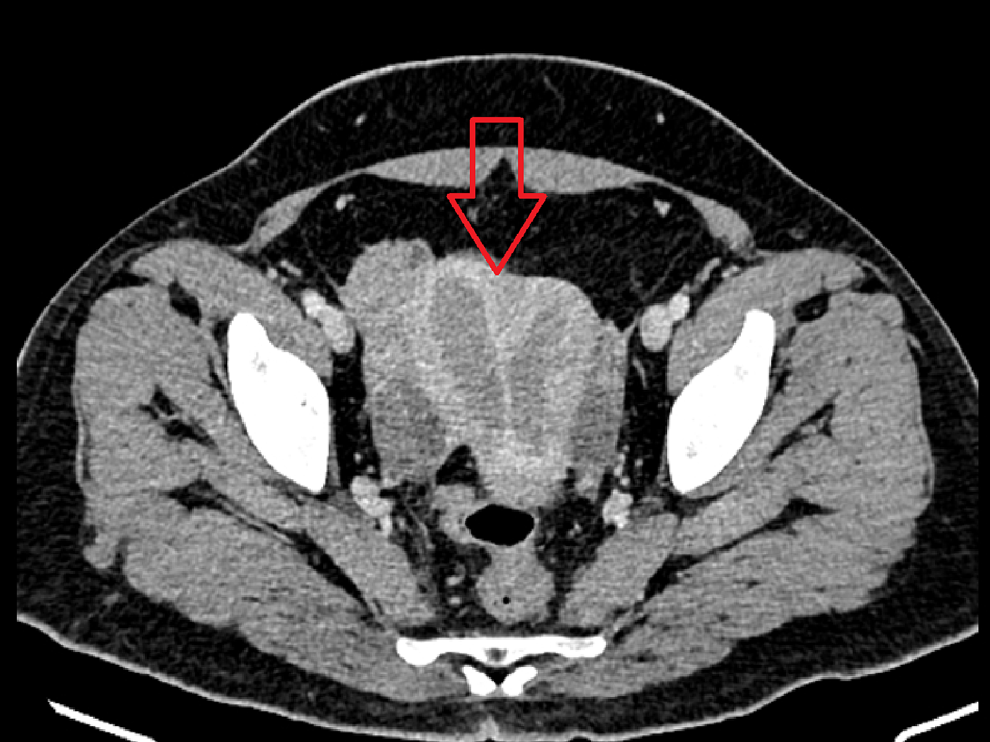
Cureus | Rare Case Report of an Endometrial Adenocarcinoma Arising in a  Complete Septate Uterus With a Double Cervix and Vagina
Cureus | Rare Case Report of an Endometrial Adenocarcinoma Arising in a Complete Septate Uterus With a Double Cervix and Vagina

Normal or Abnormal? Demystifying Uterine and Cervical Contrast Enhancement  at Multidetector CT | RadioGraphics
Normal or Abnormal? Demystifying Uterine and Cervical Contrast Enhancement at Multidetector CT | RadioGraphics
Adenomyosis of the uterus | Radiology Case | Radiopaedia.org

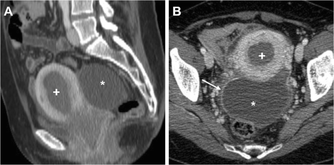
Cross-sectional imaging of acute gynaecologic disorders: CT and MRI findings  with differential diagnosis—part II: uterine emergencies and pelvic  inflammatory disease | Insights into Imaging | Full Text
Cross-sectional imaging of acute gynaecologic disorders: CT and MRI findings with differential diagnosis—part II: uterine emergencies and pelvic inflammatory disease | Insights into Imaging | Full Text

Normal or Abnormal? Demystifying Uterine and Cervical Contrast Enhancement  at Multidetector CT | RadioGraphics
Normal or Abnormal? Demystifying Uterine and Cervical Contrast Enhancement at Multidetector CT | RadioGraphics

Computed Tomography: Normal Anatomy, Imaging Techniques, and Pitfalls |  Radiology Key
Computed Tomography: Normal Anatomy, Imaging Techniques, and Pitfalls | Radiology Key

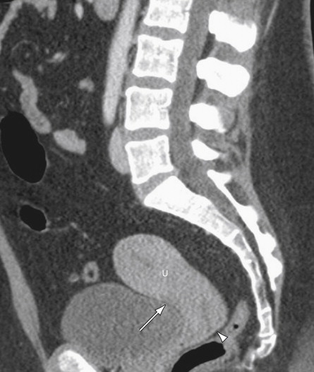
Ct scan of the abdomen reveals a uterus (U) with dilated cavity (arrow)...  | Download Scientific Diagram
Ct scan of the abdomen reveals a uterus (U) with dilated cavity (arrow)... | Download Scientific Diagram

View Image
View Image

Computed Tomography: Normal Anatomy, Imaging Techniques, and Pitfalls |  Radiology Key
Computed Tomography: Normal Anatomy, Imaging Techniques, and Pitfalls | Radiology Key

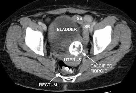
Uterine fibroids - calcified | Radiology Case | Radiopaedia.org

Exophytic Lymphoma of the Uterus Identified by 18-FDG PET Scan

CT transversal imaging of abscess formation in front of uterus. A)... |  Download Scientific Diagram
CT transversal imaging of abscess formation in front of uterus. A)... | Download Scientific Diagram

Gravid uterus | Radiology Case | Radiopaedia.org

CT scan of pelvis showing uterine hypodense mass. | Download Scientific  Diagram
CT scan of pelvis showing uterine hypodense mass. | Download Scientific Diagram

Normal or Abnormal? Demystifying Uterine and Cervical Contrast Enhancement  at Multidetector CT | RadioGraphics
Normal or Abnormal? Demystifying Uterine and Cervical Contrast Enhancement at Multidetector CT | RadioGraphics

When would you suspect fibroid uterus?

CT scan of abdomen and pelvis. Enlarged uterus with a markedly... |  Download Scientific Diagram
CT scan of abdomen and pelvis. Enlarged uterus with a markedly... | Download Scientific Diagram

Cross-sectional imaging of acute gynaecologic disorders: CT and MRI findings  with differential diagnosis—part II: uterine emergencies and pelvic  inflammatory disease | Insights into Imaging | Full Text
Cross-sectional imaging of acute gynaecologic disorders: CT and MRI findings with differential diagnosis—part II: uterine emergencies and pelvic inflammatory disease | Insights into Imaging | Full Text

CT Atlas | GLOWM
CT Atlas | GLOWM

Computed Tomography Imaging of the Acute Pelvis in Females - ScienceDirect

Gynecologic Causes of Acute Pelvic Pain: Spectrum of CT Findings |  RadioGraphics
Gynecologic Causes of Acute Pelvic Pain: Spectrum of CT Findings | RadioGraphics

Normal or Abnormal? Demystifying Uterine and Cervical Contrast Enhancement  at Multidetector CT | RadioGraphics
Normal or Abnormal? Demystifying Uterine and Cervical Contrast Enhancement at Multidetector CT | RadioGraphics

Radiological findings. A pelvic CT scan demonstrated a 5 cm uterine... |  Download Scientific Diagram
Radiological findings. A pelvic CT scan demonstrated a 5 cm uterine... | Download Scientific Diagram

Computed Tomography: Normal Anatomy, Imaging Techniques, and Pitfalls |  Radiology Key
Computed Tomography: Normal Anatomy, Imaging Techniques, and Pitfalls | Radiology Key